Pet Articles
Cat scans and MRIs

Sometimes it is easy for a veterinarian to figure out what is wrong with your pet by simple observation. Other times blood work is used to pick up problems with the thyroid glands, kidneys, or other organs. Often though, the veterinarian will decide to do further tests such as ultrasound and x-rays to look inside the body. These forms of testing are used very routinely for everything from broken bones to cancer. But what happens if none of these tests result in a diagnosis? The testing we just talked about is used to help us determine what is happening in the body. Sometimes all of the tests are negative or the diagnosis is not specific enough. This is when the veterinarian turns to different options.
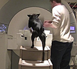
The next option might be a CT or MRI scan. Both of these options are only available at specialty practices or veterinary schools. They are very expensive, but on the other hand, they often allow veterinarians to exactly diagnose the problem. In fact, both CT and MRI are so precise that for some problems it is almost as good as looking inside the body! The CT and MRI machines are the same as those used in human hospitals. This article will give you a short introduction to the ideas and science behind these forms of testing. Then we will go through a few situations when CT and MRI are used.
“CT” stands for “computed tomography”. It is also known as a CAT scan. In order to understand CT scans, we must first do a short review of x-rays. X-rays, or “radiographs” in medical language, are created by sending a wave of high energy invisible particles through the body. These particles hit the x-ray film and record a picture. X-rays are only in black and white. The black areas are where the particles traveled easily through the body and hit the film. The white areas are where the particles were stopped inside of the body by a dense object (like bone) and didn’t hit the film, so that part of the film doesn’t turn black.
X-rays look at the whole thickness of the body and convert it to a flat image, like a camera converts a scene to a flat picture. CT scans use similar technology to x-rays but don’t put the whole thickness of the body on one picture. Instead CT can take many pictures, with each one representing a very small slice of the body. When all of those slices, like slices of a cucumber, are stacked on top of each other by a computer, you have a 3-D image of the body.
“MRI” stands for “magnetic resonance imaging”. It can also be called NMRI. MRIs were built with the same basic goal in mind as CTs- to create a 3-D image of the body. But the science behind a MRI is much different than that behind a CT scan. The MRI machine has a very large magnet. Also, every animal’s body is full of water molecules. These are two very important facts because every molecule (including water molecules) in the world has a very small magnetic field of its own. The large magnet in the MRI machine changes the direction of the magnetic fields of all the water molecules in the body. Different types of body tissues (for example, bones and organs) are affected by the magnet in different ways. These differences are detected by the MRI machine, which then creates an image.
Both CT and MRI provide great 3-D images of the body. But different problems are better diagnosed with one of the two techniques. The decision on whether to use a CT or MRI is sometimes easy if the clinic only has one of the two machines. However, in general, MRIs give clearer images of soft tissues (anything that isn’t bone) than CT scans. MRIs also do not have the potentially harmful radiation that CTs (and x-rays) need in order to produce the image. However, due to the magnet, animals with pacemakers and metal implants cannot have an MRI done.
CTs and MRIs are very detailed ways of looking inside of the body, and have been extremely useful in helping advance the quality of medicine available for pets. The science behind CTs and MRIs is very complicated. Hopefully this article has given you a brief explanation about the testing that your pet may have to go through at some point in its life.
By Ashley O’Driscoll – Pets.ca writer
3 Responses to this Article, So Far
Leave a Comment
(Additional questions? Ask them for free in our dog - cat - pet forum)

Thank youf or a very helpful description of CT & MRI imaging
if there is a choice which is better for looking for a brachial plexus tumour, a CT or and MRI
I have a rescue cat this is a Himalayan. She is around 7-8 years old and I have had her for 5 months. She began coughing a week ago which sounded very congested. I thought it was allergies. Blood work was done and everything came back normal. Xray revealed a mass in her lungs. The mass is not solid and is large 3×5. They suspect cancer. A needle aspiration cannot be done because the mass is solid. Recommendation is CT scan and surgery. Costing $6,000. Why can’t an ultrasound be done to see if cancer is in other areas before this costly surgery? What do you recommend?
I recommend you call the vet or vet tech and ask this question to them. They know your case and asking them this question should be free.
Here’s a general article on ultrasounds that may help. http://www.pets.ca/dogs/tips/dogs-cats-and-ultrasound-pet-tip-216/
For a better back and forth though, please feel free to join our forum (it’s free) and post your question there.
Good luck!